
For more information about performing a full clinical assessment to diagnose PNH, Download the Clinical Diagnostic Cheat Sheet.
High-sensitivity flow cytometrya (HSFC) is the gold standard diagnostic test for PNH1-3
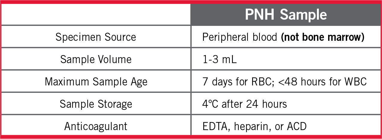
- HSFC is performed on peripheral blood—not bone marrow1,2
- Labs should perform PNH HSFC on white blood cells (WBCs) (using >1 reagent) and on red blood cells (RBCs)1,4
- Granulocytes using FLAER/CD24 combinations give most accurate estimate of PNH clone size2
- Evaluation of RBCs alone may not be accurate due to hemolysis and the dilution effect of transfusions1,4
Clear reporting is essential for appropriate clinical decisions and should include1,4:
- Clone size for each cell lineage (ie, granulocytes, monocytes, and RBCs)
- Proportion of Type II and III (percentage of GPI-deficient cells) as well as total RBCs
- Sensitivity level used (0.01% high-sensitivity analysis is ideal)
- All previous flow results in order to monitor clonal expansion
- The International PNH Interest Group (IPIG) recommends routine monitoring every 6 to 12 months of patients who have an identified clone5
aDetects PNH cells down to a 0.01% clone size.
Labs should perform PNH HSFC on RBCs, and on WBCs using >1 reagent1,4
- Evaluation of RBCs alone may underreport clone size due to hemolysis and the dilution effect of transfusions1,4
- “Routine” CD55- and/or CD59-based approaches are neither accurate nor sensitive below the 1%-4% clone size2
- Granulocytes give most accurate estimate of PNH clone size2
- FLAER/CD24 combinations recommended to detect PNH granulocytes2
- Low RBC clone size compared to WBC clone size indicative of intravascular hemolysis1,4
- The International PNH Interest Group (IPIG) recommends that high-sensitivity flow cytometry for PNH be performed5:
- After establishment of a PNH diagnosis:
- Every 6-12 months
- In patients with AA or MDS:
- At diagnosis and yearly, regardless of clone identification
- After establishment of a PNH diagnosis:
References: 1. Borowitz MJ, et al. Cytometry B Clin Cytom. 2010;78(4):211-230. 2. Sutherland DR, et al. Cytometry B Clin Cytom. 2018;94(1):23-48. 3. Sharma VR. Clin Adv Hematol Oncol. 2013;11 Suppl 13(9):2-8. 4. Illingworth A, et al. Cytometry B Clin Cytom. 2018;94(1):49-66. 5. Parker C, et al. Blood. 2005;106(12):3699-3709.